Combining computer modeling with interactive user interface to create a 3D learning tool
Gameplay Modes
QUIZ MODE
EXPLORE MODE
SLICER MODE
DTI MODE


Test your knowledge of specific brain regions.
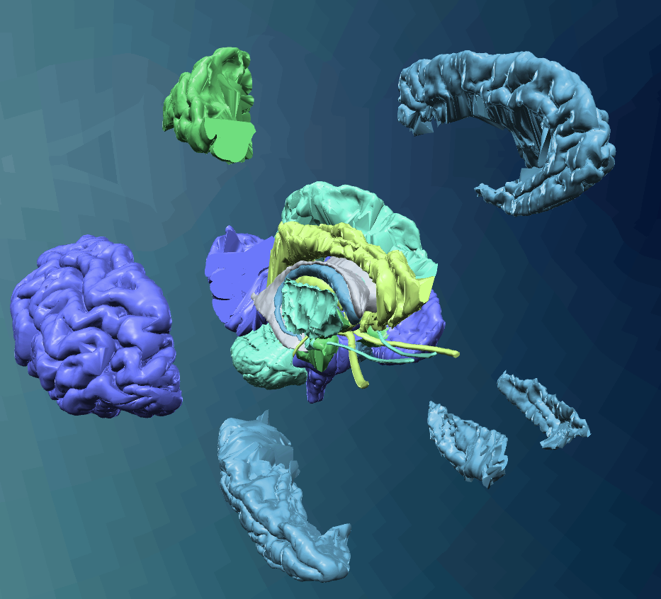
Self-guided exploration of the brain.
Axial brain slices.
Diffusion tensor imaging.
Tools to Know
ROTATE
ZOOM
SKIP


Circle around the brain with your mouse.
Zoom in and out of the brain using your mouse.
Skip questions during the quiz mode.
IDENTIFY
UNDO
MOVE


Hover over a brain part to see the name of it appear and tap to select the brain part.
Returns the last brain piece you moved.
Move brain parts around by clicking the right mouse key and then drag the section to any location.
Understanding the anatomy of complex organs such as the human brain can be a challenge for students, so there are advantages to exploring the structures in a 3-dimensional environment that provides immediate feedback as well as the ability to maneuver around the brain itself. Since 2016, the Marquette Visualization Lab has been utilizing a dataset of 3 and 7 Tesla MRI scans from the Structural Informatics Group at the University of Washington to visualize the structures of the brain in their Cave, utilizing the Unity game engine and 10 stereoscopic 3D projectors. Most MRI scans are presented as a volumetric rendering, but this dataset is unique because it was fully labeled and parcellated allowing the lab users view the structures of the brain and dissect it themselves on a device of their choice.
COVID-19 presented a new set of educational limitations, especially for courses that require students to understand such rigorous and complex information. Neuroanatomy learning techniques began with a textbook but now has evolved to include technology. One of the first for our project was the MARVL visualization studio which allowed students to view the brain but not interact with it. We then transferred the cave to the Oculus Quest, where VR abilities allowed individuals to actively manipulate the brain in a 3D manner. However, both of those versions didn't include all of the components we wanted such as immediate feedback and interactivity. This led us to development of the WebGL version which allows students to study the anatomy of the brain from most devices. Additionally, a Quiz mode was developed to enable students to not only dissect the brain and learn the anatomy, but test their knowledge of specific functions of each brain part. The programming behind this development was focused on transforming prior Oculus Quest abilities to those that would operate on a device using keyboard features. A huge benefit with the WebGL version is that the hands-on, independent, and active experience has increased student motivation to learn and overall interest in the subject.
This program uses data created and edited by John Sundsten, Associate Professor Emeritus in the Department of Biological Structure at the University of Washington, over about a 15 year period ending about 2000. These files are copyright to Structural Informatics Group, University of Washington, 2002 under the GNU Lessor Public License.